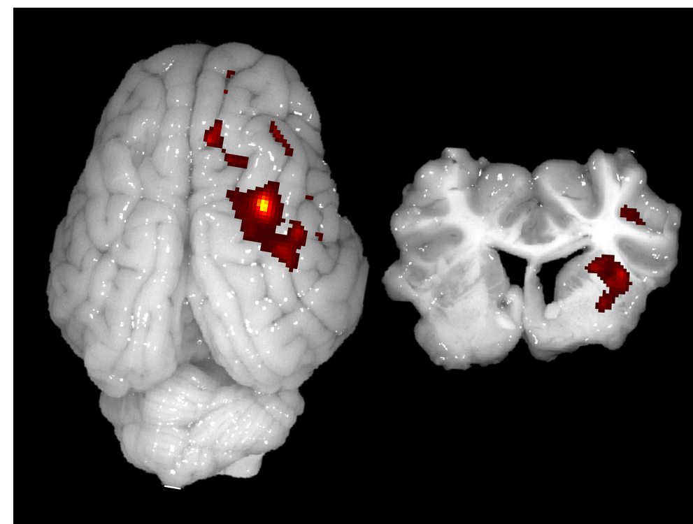
University of Georgia Core Facilities provide state-of-the-art equipment and services to the TNRR Laboratory. These Core Facilities make use of highly specialized scientific equipment, diagnostic tools, adaptable prototyping processes, and mechanical production shops.
The Proteomics and Mass Spectrometry Core Facility is equipped with an ThermoScientific Orbitrap Elite mass spectrometer for high resolution and high mass accuracy analysis. It is coupled with a nano HPLC, increasing its capacity to analyze more complex protein mixtures. The facility also has a Bruker Autoflex MALDI for quick analysis of tryptic digests of pure proteins. The facility offers in-gel digestion and subsequence analysis for protein identification. The facility also has an in-house version of Mascot that provides customers with the option of loading a database to search for protein identification.
The Coverdell Rodent Vivarium provides two Perkin Elmer in vivo imaging systems are available to meet the TNRR Laboratory’s live animal imaging needs. The IVIS Lumina II is an expandable, sensitive imaging system that allows both fluorescent and bioluminescent imaging in vivo. It can accommodate rodents, petri dishes, microtiter plates, and isolated organ systems for in vitro or ex vivo imaging. Maestro 2 enables high sensitivity in vivo fluorescence imaging of small animals. This system also generates time-based kinetic images as well as videos of fluorescent reagents and labeled antibodies, thus providing quantitative data on the temporal biodistribution of fluorescent markers. This kinetic imaging data can be used to generate all-optical anatomic rodent organ maps as a part of body-compartment modeling, pK value calculations or rate of uptake, and washout determinations. For more information please refer to https://ctegd.uga.edu/resources/animal-imaging/.

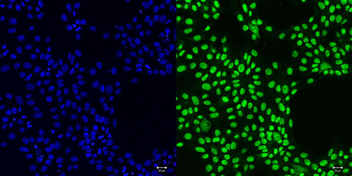
The Biomedical Microscopy Core provides access to confocal, deconvolution, light sheet, super resolution and other optical microscope systems that are useful for multicolor imaging of live and fixed cells and tissue samples, and high-content screening. This state-of-the-art microscopy facility serves UGA and the TNRR by providing microscopy related expertise, training and assistance for advancing projects on various model organisms.

For more information on UGA’s Core Facilities please refer to https://research.uga.edu/core-facilities/.