Progenitor/stem cell source of taste bud cells
Our recent findings revealed distinct progenitor sources for early and mature taste buds and a previously unrecognized stem cell source for taste buds. Mapping lineage of E11.0 Shh+ epithelium of the tongue rudiment with Shh-CreERT2/RFP mice demonstrated that the early taste buds and the surrounding lingual epithelium are from the same population of progenitors, i.e., Shh+ epithelial cells of the tongue primordium. In combination with previous reports, we propose that Shh+Krt8+ cells in the homogeneous epithelium of tongue primordium at early embryonic stages (E11.0-11.5) are progenitors for taste papilla and taste bud cells, and non-gustatory cells surrounding papillae, including K14+ basal epithelial cells which will eventually contribute to the cell renewal of mature taste buds.

Recently, we found that SOX10+ cells in the von Ebner’s glands give rise to taste bud cells during maturation and maintenance, which is a novel concept in the field and opens new avenues of study regarding taste bud formation and homeostasis

Requirement of tongue mesenchyme for the proper tongue development and taste papilla differentiation
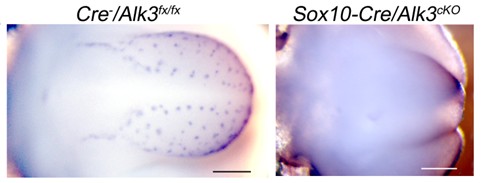
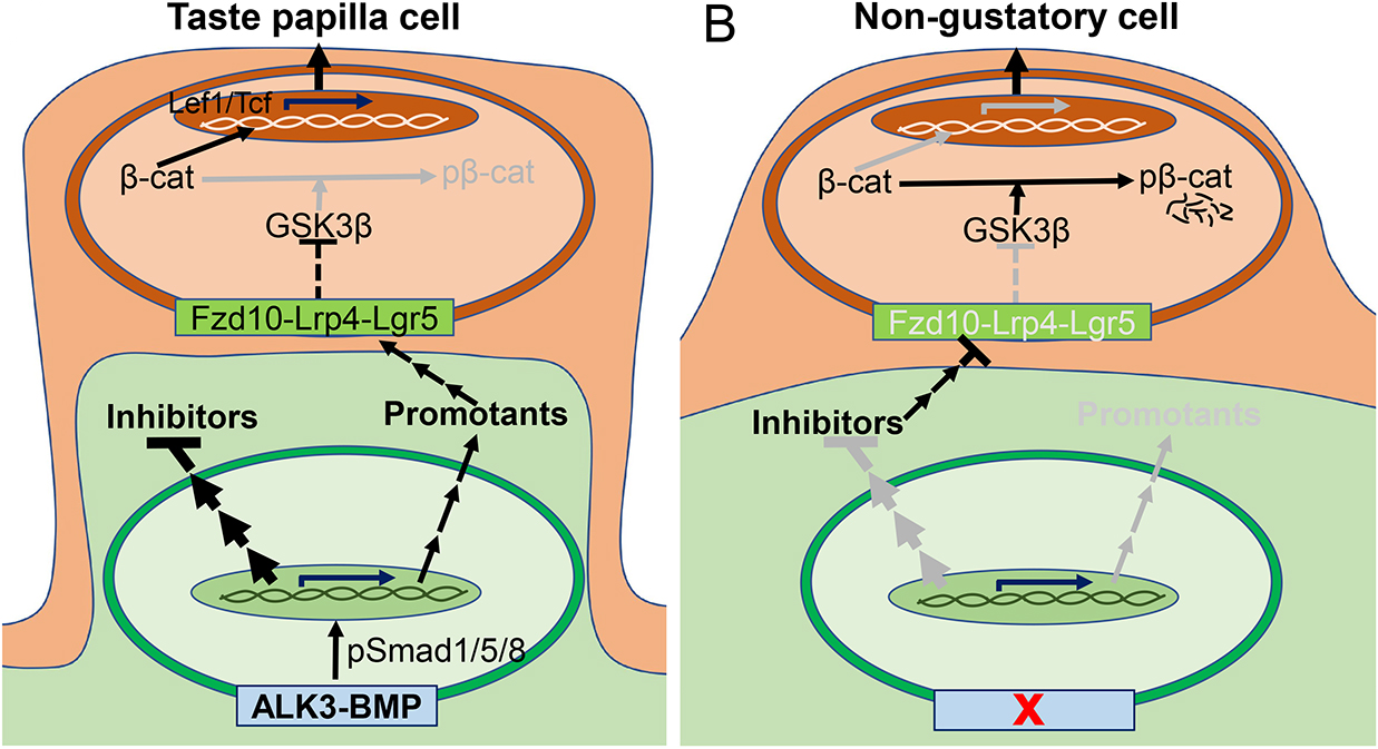
Taste papillae are specialized organs, each of which comprises an epithelial wall hosting taste buds and a core of mesenchymal tissue. Using mesenchyme-specific genomic modifications of receptors for bone morphogenetic protein (BMP) (constitutive activation of Alk2) or neurofibromatosis 2 (Nf2) by Wnt1-Cre and Sox10-Cre, we have demonstrated that molecular signaling pathways in the tongue mesenchyme regulates the proper development of tongue shape and size in a region-specific manner. Recently, we reported that during early taste papilla development in mouse embryos, bone morphogenetic protein (BMP) signaling mediated by type 1 receptor ALK3 in the tongue mesenchyme is required for epithelial Wnt/β-catenin activity and taste papilla differentiation. Our findings demonstrate for the first time the requirement of tongue mesenchyme in taste papilla cell differentiation.
Establishment of a mouse model mimicking human disease with taste bud loss
Familial Dysautonomia (FD) is a rare neurodevelopmental and neurodegenerative disorder that affects the sympathetic and sensory nervous system. FD patients have ELP1 reduction and functional deficits of the peripheral nervous system (PNS) including taste loss. We have successfully established a disease model (tissue-specific knockout of Elp1) to study the defects of nervous system including loss of taste buds.
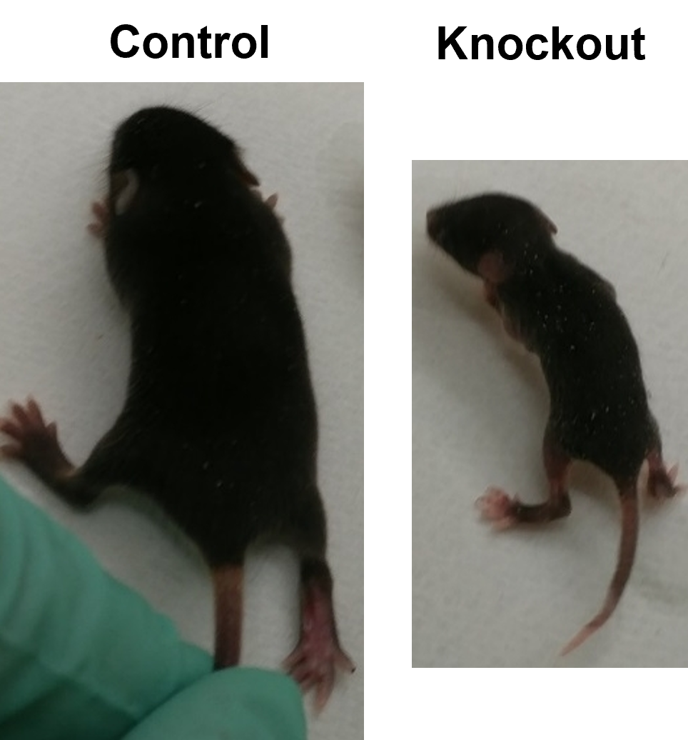
In collaboration with Dr. Nadja Zeltner (CMM, UGA) and Frances Lefcort (Montana State University), we have found that FD sympathetic neurons (symNs) are intrinsically hyperactive in vitro and ex vivo, which may be the cause of neuronal death/degeneration. Importantly, the Elp1 cKO mice depicts taste bud defects and a reduction of taste sensitivity which provides a model for mechanistic studies on taste bud formation and maintenance.